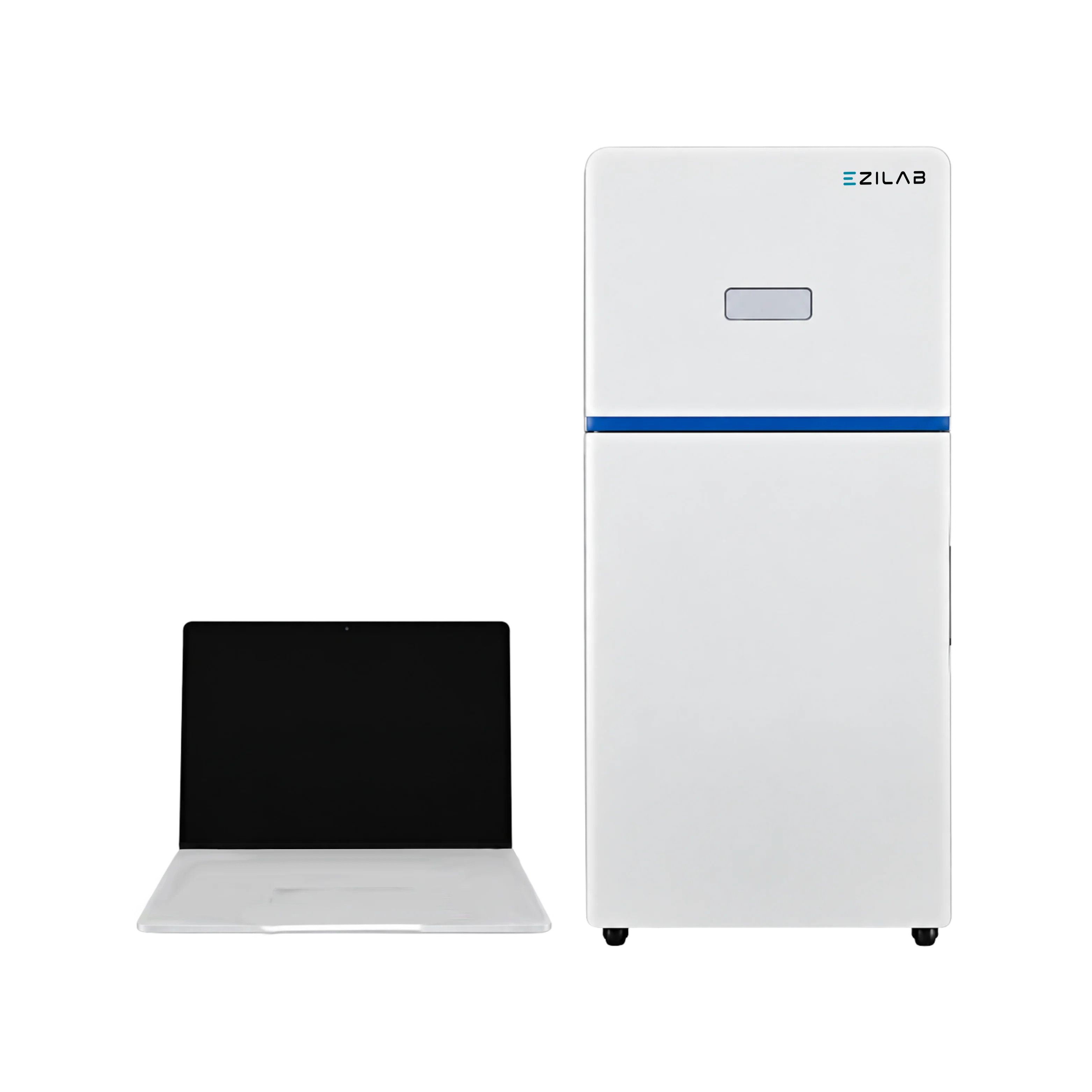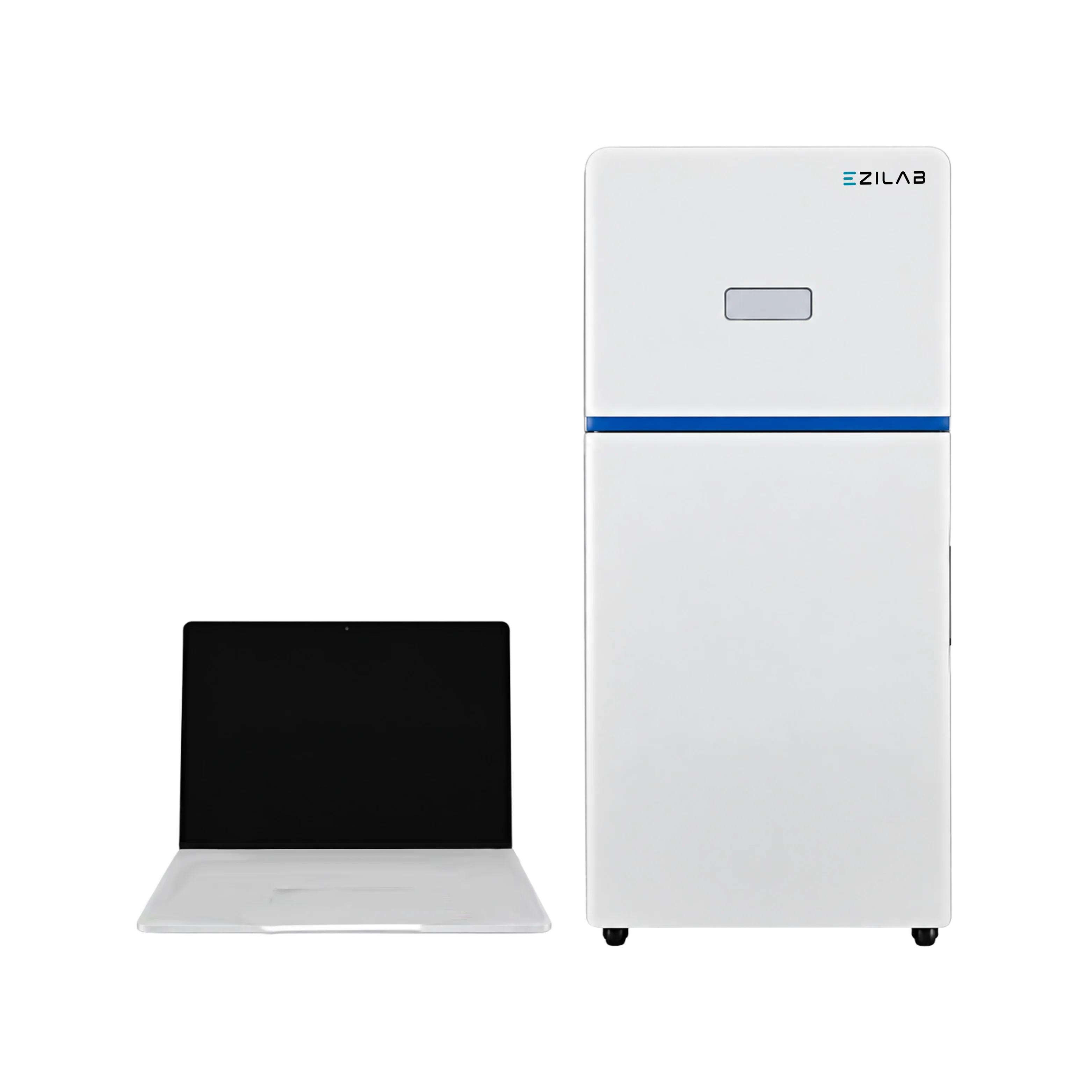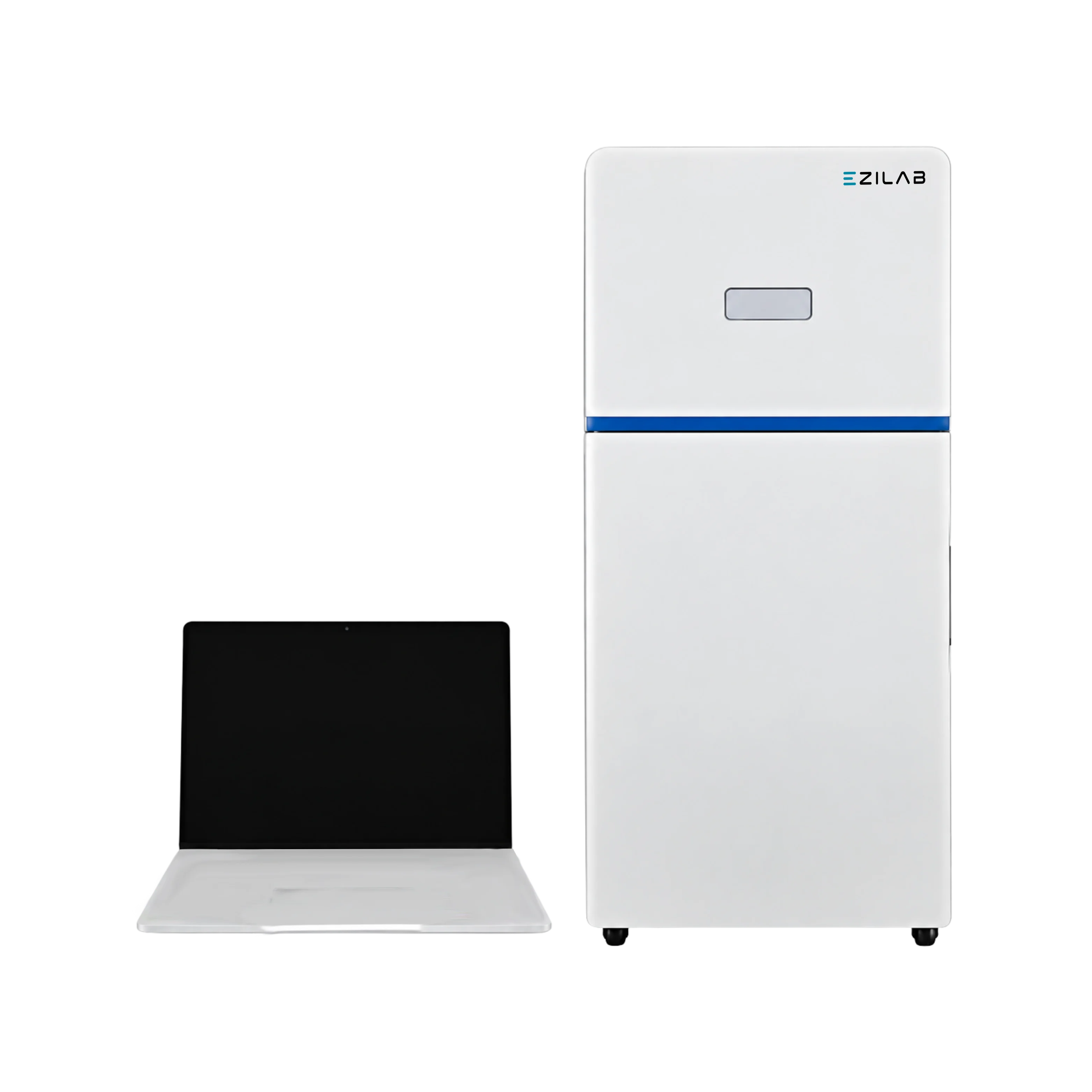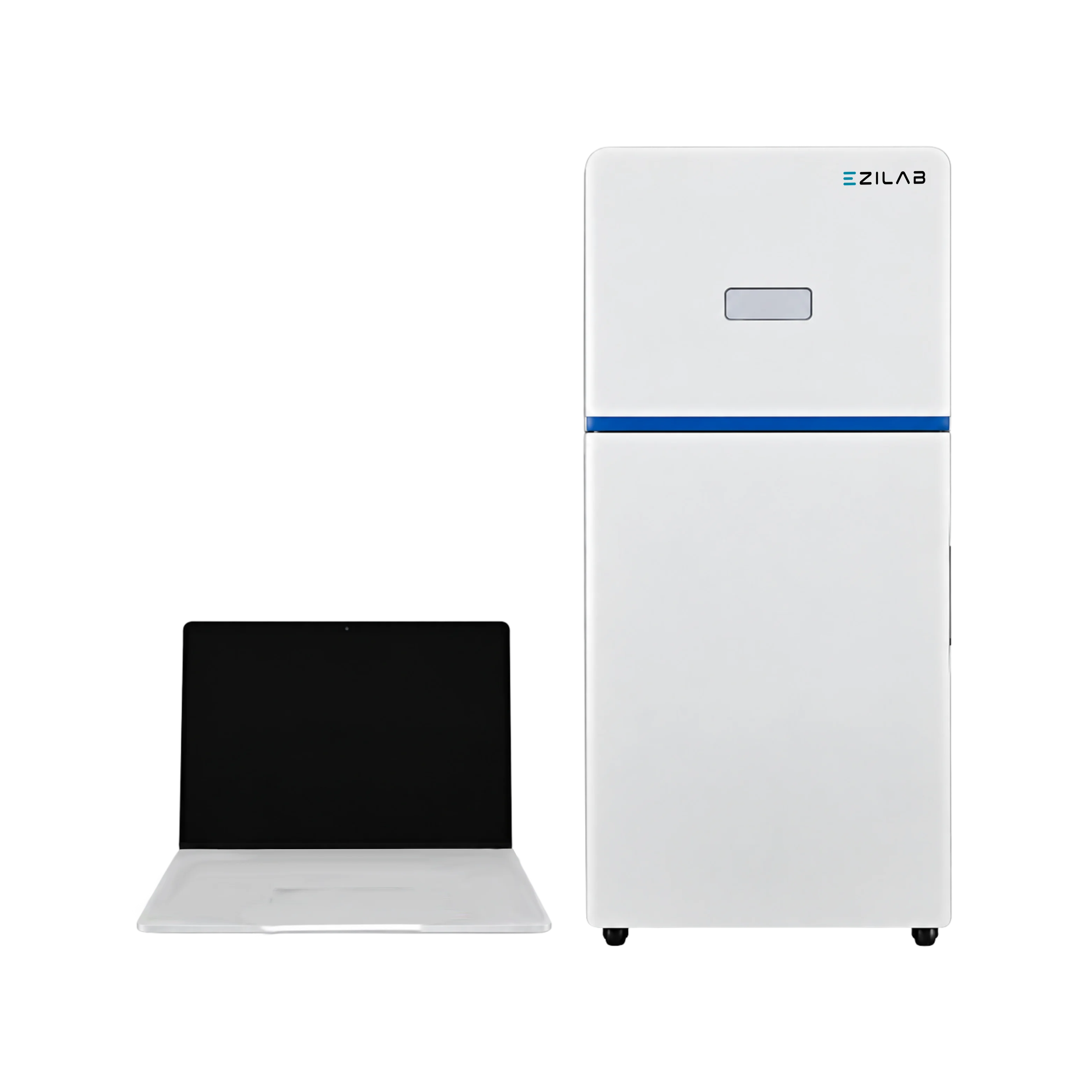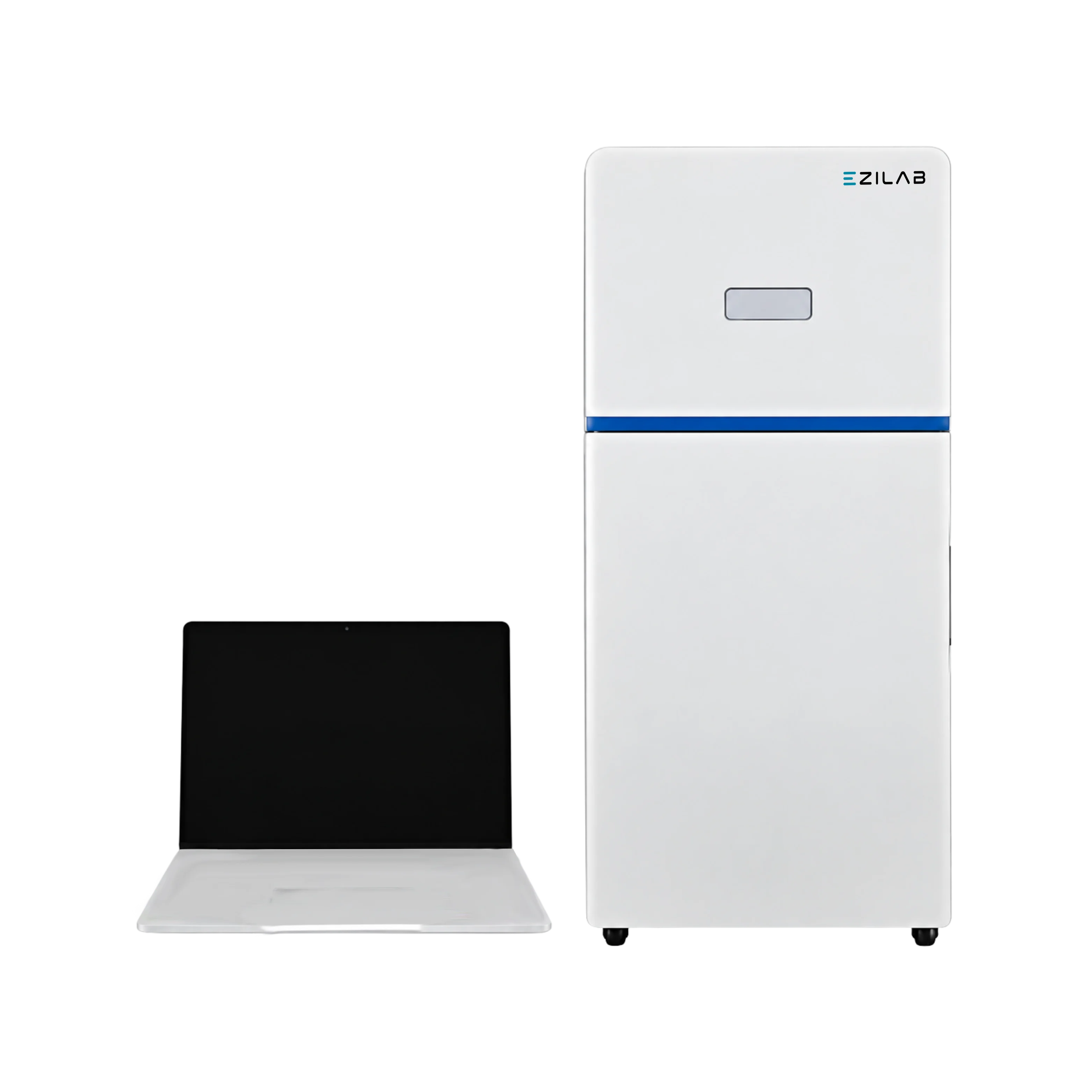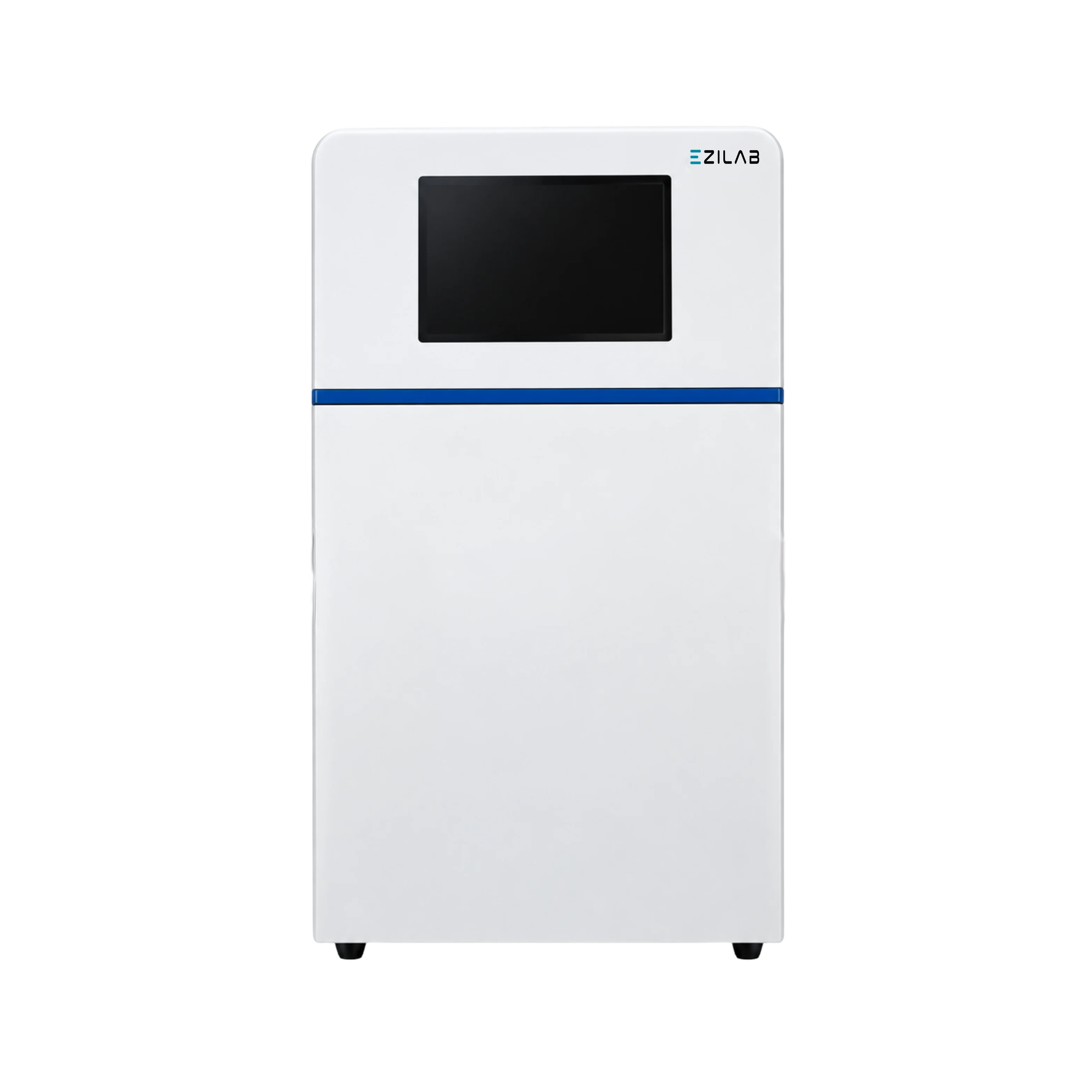
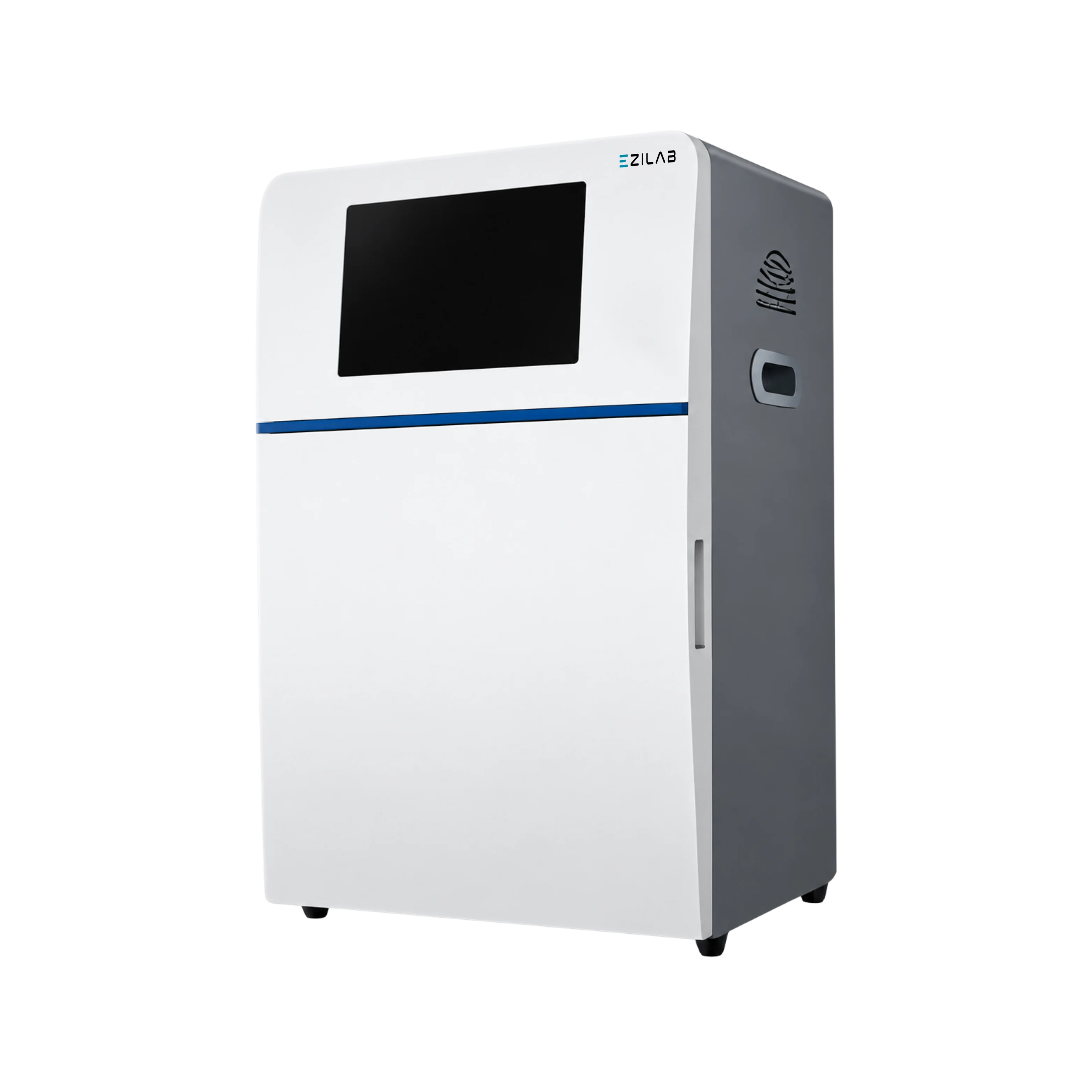
EziLab offers the Gel Documentation Imaging System EZL-GD133, a gel imaging unit that captures DNA, RNA and protein gels on a 5 megapixel camera driven by an embedded computer and touchscreen. The 2592 by 1944 sensor delivers 70% quantum efficiency, 5.1 e- read noise, 70.1 dB signal-to-noise and 1 e- per pixel per second dark current. An 8 position filter wheel matches the stain, UV, blue and white light transilluminators switch in software, automatic exposure and pixel binning set themselves, and workflows store as protocols. On-board tools crop, rotate, invert, wipe background and compute optical density, while the RIM-molded polymer cabinet keeps the 31 kg darkroom light-tight. Molecular biology laboratories, genomics core facilities, protein chemistry groups, microbiology departments, pharmaceutical QC teams and university teaching labs use this gel documentation system to record and quantify electrophoresis results. Typical work includes agarose and SDS-PAGE gel capture, Western blot chemiluminescence, ELISA plate imaging, colony counting on Petri dishes and TLC plate documentation. Choose it over a UV cabinet and phone camera when band intensities must be quantified and archived. Ask whether raw images export as 16-bit TIFF: densitometry on compressed JPEG files is hard to defend in a data review. Imaging System holds the other imaging families, with Gel Doc Imaging System as the electrophoresis group.
| Camera | Embedded industry computer with touch screen |
|---|---|
| Resolution | 5.0 Megapixels, 2592×1944 |
| Quantum Efficiency | ≥ 70% |
| Read Out Noise | 5.1e-RMS |
| SNR | 70.1db |
| A/D | 1e-/pixel/sec.@25°C |
| Dark Current | 1e-/pixel/sec.@25°C |
| Filter Wheel | 8 sockets filter wheel |
| Product Dimension (W×D×H) | 430×380×690 mm |
| Outer Pack Dimension (W×D×H) | 550×490×760 mm |
| Gross Weight | 31 kg |
| Feature Area | Description |
| Illumination | A UV trans illuminator is joined by blue and white light trans illuminators for different stains. |
| Operation | An embedded computer with a touchscreen offers switchable operation modes. |
| Exposure | Automatic exposure and automatic access of capture parameters work with saved protocols. |
| Image Tools | Cropping, rotation, colour inversion and a wiping background mode tidy the final image. |
| Analysis | Optical density calculation supports quantitative analysis of bands. |